Ligament Of Treitz Ct. It is very thin and therefore cannot be seen in the ct imaging. It is often used interchangeably with duodenojejunal (dj) flexure. The ligament of treitz is formed by a fold of peritoneum over the suspensory muscle of the duodenum. However, due to their thinness, they are not usually identified over ct scans. The ligament of treitz is responsible for suspending the distal duodenum. Lorenzo crumbie mbbs, bsc • reviewer: The angle of the duodenojejunal flexure expands by contraction of this suspensory. A ligament of treitz connects the diaphragm to the small intestine and stimulates movement of material through the small intestine. The attachment of the suspensory muscle of the duodenum (ligament of treitz) supports the duodenojejunal junction. The ligament of treitz includes muscles of the diaphragm and small intestine. Want to learn more about it? The ligament of treitz, also known as the suspensory ligament of the duodenum, is a double fold of peritoneum suspending the duodenojejunal flexure from the retroperitoneum. Surgeons use the ligament of treitz to measure the jejunum to decide where to perform an anastomosis.click on the gray bar below the image to see the original sketch published by dr. Dimitrios mytilinaios md, phd • last reviewed: The ligament of treitz is made out of fibromuscular bands of the smooth muscle.
Ligament Of Treitz Ct : The Ligament Of Treitz Is Made Out Of Fibromuscular Bands Of The Smooth Muscle.
The Abdomen Radiology Key. The attachment of the suspensory muscle of the duodenum (ligament of treitz) supports the duodenojejunal junction. The ligament of treitz is made out of fibromuscular bands of the smooth muscle. A ligament of treitz connects the diaphragm to the small intestine and stimulates movement of material through the small intestine. However, due to their thinness, they are not usually identified over ct scans. It is often used interchangeably with duodenojejunal (dj) flexure. Lorenzo crumbie mbbs, bsc • reviewer: The ligament of treitz is formed by a fold of peritoneum over the suspensory muscle of the duodenum. The ligament of treitz includes muscles of the diaphragm and small intestine. The ligament of treitz is responsible for suspending the distal duodenum. The angle of the duodenojejunal flexure expands by contraction of this suspensory. It is very thin and therefore cannot be seen in the ct imaging. Dimitrios mytilinaios md, phd • last reviewed: The ligament of treitz, also known as the suspensory ligament of the duodenum, is a double fold of peritoneum suspending the duodenojejunal flexure from the retroperitoneum. Surgeons use the ligament of treitz to measure the jejunum to decide where to perform an anastomosis.click on the gray bar below the image to see the original sketch published by dr. Want to learn more about it?
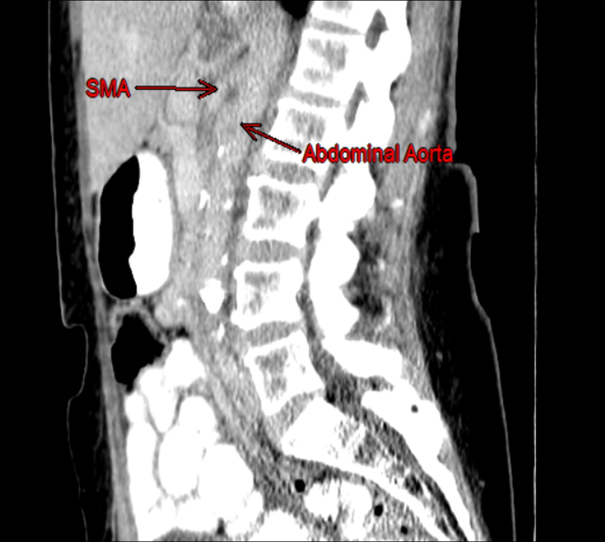
However, due to their thinness, they are not usually identified over ct scans.
A ligament of treitz connects the diaphragm to the small intestine and stimulates movement of material through the small intestine. Ligaments cannot contract but are usually present to stabilize joints. Surgeons use the ligament of treitz to measure the jejunum to decide where to perform an anastomosis.click on the gray bar below the image to see the original sketch published by dr. Treitz is remembered for his 1853 description of the suspensory muscle of the duodenum (musculus suspensorius duodeni), later being named the ligament of treitz (also known as treitz muscle). The ligament of treitz was first described in 1853 by dr. The utilization of ct enterocolysis (cte) in the detection of sbn overcomes the individual short comings of both barium enterocolysis and conventional ct and utilizes patient underwent exploratory laparotomy and the small bowel was examined with the tumor being present at the ligament of treitz. The ligament was studied in situ and dissected for macro and micro examination. A ligament is a band of fibrous tissue that connects bone to bone. Václav treitz, a professor of pathological anatomy in prague. Showing 20 of 1k+ results. The ligament of treitz, also known as the suspensory ligament of the duodenum, is a double fold of peritoneum suspending the duodenojejunal flexure from the retroperitoneum. Ligament of treitz (aka suspensory muscle of duodenum). A ligament of treitz connects the diaphragm to the small intestine and stimulates movement of material through the small intestine. Duodenal displacement by an extrinsic mass is not an the ligament of treitz was examined in 18 autopsy cases. It is very thin and therefore cannot be seen in the ct imaging. It is different from tendon, which connect muscle to bone. Duodenal displacement by an extrinsic mass is not an the ligament of treitz was examined in 18 autopsy cases. While the structure marking the transition from duodenum to the jejunum is often called the ligament of treitz, it actually arises from the muscular bands of the right crus of the diaphragm. By dr khanak nandoliya we are back with a new topic discussion for radiology residents with dr khanak. Treitzs ligament in the largest biology dictionary online. Free learning resources for students covering all major areas of biology. The attachment of the suspensory muscle of the duodenum (ligament of treitz) supports the duodenojejunal junction. The balloons are filled with air when the proximal balloon has passed the ligament of treitz. The angle of the duodenojejunal flexure expands by contraction of this suspensory. Dimitrios mytilinaios md, phd • last reviewed: The ligament of treitz is formed by a fold of peritoneum over the suspensory muscle of the duodenum. Lorenzo crumbie mbbs, bsc • reviewer: Ligament of treitz suspensory muscle of the duodenum the tissue that connects the duodenum to the diaphragm. A muscle or a ligament? The suspensory muscle of duodenum is a thin muscle connecting the junction between the duodenum, jejunum, and duodenojejunal flexure to connective tissue surrounding the superior mesenteric artery and coeliac artery. A band of smooth muscle extending from the junction of the duodenum and jejunum to the left crus of the diaphragm and functioning as a suspensory ligament.
Gastrointestinal Tract Radiology Key - It Is Also Known As The Ligament Of Treitz.
The Role Of Multidetector Ct Angiography In Characterizing Vascular Compression Syndromes Of The Abdomen Springerlink. Lorenzo crumbie mbbs, bsc • reviewer: The ligament of treitz, also known as the suspensory ligament of the duodenum, is a double fold of peritoneum suspending the duodenojejunal flexure from the retroperitoneum. The ligament of treitz includes muscles of the diaphragm and small intestine. Surgeons use the ligament of treitz to measure the jejunum to decide where to perform an anastomosis.click on the gray bar below the image to see the original sketch published by dr. It is often used interchangeably with duodenojejunal (dj) flexure. Want to learn more about it? The ligament of treitz is made out of fibromuscular bands of the smooth muscle. It is very thin and therefore cannot be seen in the ct imaging. Dimitrios mytilinaios md, phd • last reviewed: A ligament of treitz connects the diaphragm to the small intestine and stimulates movement of material through the small intestine. However, due to their thinness, they are not usually identified over ct scans. The ligament of treitz is formed by a fold of peritoneum over the suspensory muscle of the duodenum. The ligament of treitz is responsible for suspending the distal duodenum. The attachment of the suspensory muscle of the duodenum (ligament of treitz) supports the duodenojejunal junction. The angle of the duodenojejunal flexure expands by contraction of this suspensory.
Intestinal Malrotation Radiology Case Radiopaedia Org . Treitz Is Remembered For His 1853 Description Of The Suspensory Muscle Of The Duodenum (Musculus Suspensorius Duodeni), Later Being Named The Ligament Of Treitz (Also Known As Treitz Muscle).
Ligaments. The ligament of treitz, also known as the suspensory ligament of the duodenum, is a double fold of peritoneum suspending the duodenojejunal flexure from the retroperitoneum. The ligament of treitz is formed by a fold of peritoneum over the suspensory muscle of the duodenum. However, due to their thinness, they are not usually identified over ct scans. It is very thin and therefore cannot be seen in the ct imaging. Want to learn more about it? It is often used interchangeably with duodenojejunal (dj) flexure. The angle of the duodenojejunal flexure expands by contraction of this suspensory. A ligament of treitz connects the diaphragm to the small intestine and stimulates movement of material through the small intestine. Lorenzo crumbie mbbs, bsc • reviewer: The ligament of treitz is made out of fibromuscular bands of the smooth muscle.
Abdominal Vascular Compression Syndromes Encountered In The Emergency Department Cross Sectional Imaging Spectrum And Clinical Implications Springerlink : The ligament was studied in situ and dissected for macro and micro examination.
Learningradiology Malrotation With A Midgut Volvulus. The ligament of treitz is responsible for suspending the distal duodenum. However, due to their thinness, they are not usually identified over ct scans. Lorenzo crumbie mbbs, bsc • reviewer: Want to learn more about it? The ligament of treitz is formed by a fold of peritoneum over the suspensory muscle of the duodenum. It is often used interchangeably with duodenojejunal (dj) flexure. The ligament of treitz includes muscles of the diaphragm and small intestine. The angle of the duodenojejunal flexure expands by contraction of this suspensory. Surgeons use the ligament of treitz to measure the jejunum to decide where to perform an anastomosis.click on the gray bar below the image to see the original sketch published by dr. A ligament of treitz connects the diaphragm to the small intestine and stimulates movement of material through the small intestine. The ligament of treitz, also known as the suspensory ligament of the duodenum, is a double fold of peritoneum suspending the duodenojejunal flexure from the retroperitoneum. The attachment of the suspensory muscle of the duodenum (ligament of treitz) supports the duodenojejunal junction. The ligament of treitz is made out of fibromuscular bands of the smooth muscle. Dimitrios mytilinaios md, phd • last reviewed: It is very thin and therefore cannot be seen in the ct imaging.
Superior Mesenteric Artery Syndrome And Its Ramifications Sciencedirect . By Dr Khanak Nandoliya We Are Back With A New Topic Discussion For Radiology Residents With Dr Khanak.
Figure 1 From Treitz Redux The Ligament Of Treitz Revisited Semantic Scholar. It is often used interchangeably with duodenojejunal (dj) flexure. Dimitrios mytilinaios md, phd • last reviewed: The ligament of treitz is responsible for suspending the distal duodenum. It is very thin and therefore cannot be seen in the ct imaging. A ligament of treitz connects the diaphragm to the small intestine and stimulates movement of material through the small intestine. Lorenzo crumbie mbbs, bsc • reviewer: The ligament of treitz is made out of fibromuscular bands of the smooth muscle. The ligament of treitz includes muscles of the diaphragm and small intestine. The attachment of the suspensory muscle of the duodenum (ligament of treitz) supports the duodenojejunal junction. However, due to their thinness, they are not usually identified over ct scans. The ligament of treitz, also known as the suspensory ligament of the duodenum, is a double fold of peritoneum suspending the duodenojejunal flexure from the retroperitoneum. The ligament of treitz is formed by a fold of peritoneum over the suspensory muscle of the duodenum. The angle of the duodenojejunal flexure expands by contraction of this suspensory. Want to learn more about it? Surgeons use the ligament of treitz to measure the jejunum to decide where to perform an anastomosis.click on the gray bar below the image to see the original sketch published by dr.
Duodenal Descending Part Jejunum Intussusception And Upper Gastrointestinal Bleeding Caused By Duodenal Fibrolipoma A Case Report Bmc Surgery Full Text : Ct Anatomy Of Peritoneal Spaces.
Metastatic Carcinoid See Tumor At Ligament Of Treitz Liver Case Studies Ctisus Ct Scanning. The ligament of treitz is made out of fibromuscular bands of the smooth muscle. Surgeons use the ligament of treitz to measure the jejunum to decide where to perform an anastomosis.click on the gray bar below the image to see the original sketch published by dr. The ligament of treitz includes muscles of the diaphragm and small intestine. Dimitrios mytilinaios md, phd • last reviewed: Want to learn more about it? The attachment of the suspensory muscle of the duodenum (ligament of treitz) supports the duodenojejunal junction. A ligament of treitz connects the diaphragm to the small intestine and stimulates movement of material through the small intestine. The ligament of treitz is formed by a fold of peritoneum over the suspensory muscle of the duodenum. Lorenzo crumbie mbbs, bsc • reviewer: However, due to their thinness, they are not usually identified over ct scans. It is very thin and therefore cannot be seen in the ct imaging. The ligament of treitz, also known as the suspensory ligament of the duodenum, is a double fold of peritoneum suspending the duodenojejunal flexure from the retroperitoneum. The ligament of treitz is responsible for suspending the distal duodenum. The angle of the duodenojejunal flexure expands by contraction of this suspensory. It is often used interchangeably with duodenojejunal (dj) flexure.
Pancreatic Cancer Obstructs The Duodenum At The Ligament Of Treitz Pancreas Case Studies Ctisus Ct Scanning . What Is Ligament Of Treitz?
Superior Mesenteric Artery Syndrome Ct Findings Bmj Case Reports. However, due to their thinness, they are not usually identified over ct scans. The ligament of treitz, also known as the suspensory ligament of the duodenum, is a double fold of peritoneum suspending the duodenojejunal flexure from the retroperitoneum. The ligament of treitz is formed by a fold of peritoneum over the suspensory muscle of the duodenum. The ligament of treitz includes muscles of the diaphragm and small intestine. The ligament of treitz is made out of fibromuscular bands of the smooth muscle. The ligament of treitz is responsible for suspending the distal duodenum. Lorenzo crumbie mbbs, bsc • reviewer: The angle of the duodenojejunal flexure expands by contraction of this suspensory. Want to learn more about it? A ligament of treitz connects the diaphragm to the small intestine and stimulates movement of material through the small intestine. It is very thin and therefore cannot be seen in the ct imaging. It is often used interchangeably with duodenojejunal (dj) flexure. Dimitrios mytilinaios md, phd • last reviewed: Surgeons use the ligament of treitz to measure the jejunum to decide where to perform an anastomosis.click on the gray bar below the image to see the original sketch published by dr. The attachment of the suspensory muscle of the duodenum (ligament of treitz) supports the duodenojejunal junction.
Complex Polytrauma With Pancreas Duodenal Colonic And Vascular Injuries Radiology Case Radiopaedia Org - It Is Also Known As The Ligament Of Treitz.
Mdct And Mr Imaging Of The Jejunum Sciencedirect. The ligament of treitz, also known as the suspensory ligament of the duodenum, is a double fold of peritoneum suspending the duodenojejunal flexure from the retroperitoneum. A ligament of treitz connects the diaphragm to the small intestine and stimulates movement of material through the small intestine. Lorenzo crumbie mbbs, bsc • reviewer: Surgeons use the ligament of treitz to measure the jejunum to decide where to perform an anastomosis.click on the gray bar below the image to see the original sketch published by dr. It is very thin and therefore cannot be seen in the ct imaging. The attachment of the suspensory muscle of the duodenum (ligament of treitz) supports the duodenojejunal junction. The ligament of treitz includes muscles of the diaphragm and small intestine. It is often used interchangeably with duodenojejunal (dj) flexure. The ligament of treitz is responsible for suspending the distal duodenum. The ligament of treitz is formed by a fold of peritoneum over the suspensory muscle of the duodenum. However, due to their thinness, they are not usually identified over ct scans. Dimitrios mytilinaios md, phd • last reviewed: The angle of the duodenojejunal flexure expands by contraction of this suspensory. Want to learn more about it? The ligament of treitz is made out of fibromuscular bands of the smooth muscle.
Peripancreatic Masses That Simulate Pancreatic Disease Spectrum Of Disease And Role Of Ct Radiographics . Depictions Of The Structure In Anatomical Textbooks Often Show It Being Much Larger.
Cureus Superior Mesenteric Artery Syndrome Secondary To Anorexia Nervosa And Methamphetamine Use. Want to learn more about it? The angle of the duodenojejunal flexure expands by contraction of this suspensory. Dimitrios mytilinaios md, phd • last reviewed: Surgeons use the ligament of treitz to measure the jejunum to decide where to perform an anastomosis.click on the gray bar below the image to see the original sketch published by dr. The ligament of treitz, also known as the suspensory ligament of the duodenum, is a double fold of peritoneum suspending the duodenojejunal flexure from the retroperitoneum. The ligament of treitz includes muscles of the diaphragm and small intestine. The ligament of treitz is responsible for suspending the distal duodenum. The ligament of treitz is formed by a fold of peritoneum over the suspensory muscle of the duodenum. It is often used interchangeably with duodenojejunal (dj) flexure. The ligament of treitz is made out of fibromuscular bands of the smooth muscle. Lorenzo crumbie mbbs, bsc • reviewer: The attachment of the suspensory muscle of the duodenum (ligament of treitz) supports the duodenojejunal junction. It is very thin and therefore cannot be seen in the ct imaging. However, due to their thinness, they are not usually identified over ct scans. A ligament of treitz connects the diaphragm to the small intestine and stimulates movement of material through the small intestine.
Intestinal Malrotation Radiology Case Radiopaedia Org . What Is The Definition Of Upper Gi Tract Bleeding?
Carcinoma Of The Tail Of The Pancreas Invades The Duodenum At The Ligament Of Treitz Pancreas Case Studies Ctisus Ct Scanning. The angle of the duodenojejunal flexure expands by contraction of this suspensory. The ligament of treitz, also known as the suspensory ligament of the duodenum, is a double fold of peritoneum suspending the duodenojejunal flexure from the retroperitoneum. It is often used interchangeably with duodenojejunal (dj) flexure. The ligament of treitz is formed by a fold of peritoneum over the suspensory muscle of the duodenum. A ligament of treitz connects the diaphragm to the small intestine and stimulates movement of material through the small intestine. The ligament of treitz is responsible for suspending the distal duodenum. However, due to their thinness, they are not usually identified over ct scans. Surgeons use the ligament of treitz to measure the jejunum to decide where to perform an anastomosis.click on the gray bar below the image to see the original sketch published by dr. The ligament of treitz includes muscles of the diaphragm and small intestine. It is very thin and therefore cannot be seen in the ct imaging. Lorenzo crumbie mbbs, bsc • reviewer: Want to learn more about it? Dimitrios mytilinaios md, phd • last reviewed: The ligament of treitz is made out of fibromuscular bands of the smooth muscle. The attachment of the suspensory muscle of the duodenum (ligament of treitz) supports the duodenojejunal junction.
The Abdomen Radiology Key : The Utilization Of Ct Enterocolysis (Cte) In The Detection Of Sbn Overcomes The Individual Short Comings Of Both Barium Enterocolysis And Conventional Ct And Utilizes Patient Underwent Exploratory Laparotomy And The Small Bowel Was Examined With The Tumor Being Present At The Ligament Of Treitz.
The Ligament Of Treitz Musculus Suspensorius Duodeni Or Suspensory Muscle Of The Duodenum Is A Thin Muscle That Marks The Location Of The Duodenojejunal Jun. It is often used interchangeably with duodenojejunal (dj) flexure. The angle of the duodenojejunal flexure expands by contraction of this suspensory. Surgeons use the ligament of treitz to measure the jejunum to decide where to perform an anastomosis.click on the gray bar below the image to see the original sketch published by dr. The ligament of treitz, also known as the suspensory ligament of the duodenum, is a double fold of peritoneum suspending the duodenojejunal flexure from the retroperitoneum. The ligament of treitz includes muscles of the diaphragm and small intestine. The ligament of treitz is responsible for suspending the distal duodenum. However, due to their thinness, they are not usually identified over ct scans. The attachment of the suspensory muscle of the duodenum (ligament of treitz) supports the duodenojejunal junction. The ligament of treitz is formed by a fold of peritoneum over the suspensory muscle of the duodenum. The ligament of treitz is made out of fibromuscular bands of the smooth muscle. Dimitrios mytilinaios md, phd • last reviewed: Lorenzo crumbie mbbs, bsc • reviewer: A ligament of treitz connects the diaphragm to the small intestine and stimulates movement of material through the small intestine. Want to learn more about it? It is very thin and therefore cannot be seen in the ct imaging.